Revolutionizing AI in Medical Imaging
The central development is this: Artificial intelligence is rapidly transforming healthcare, yet one significant hurdle remains: access to vast, diverse, and clean clinical data, especially in sensitive areas like neuroimaging. Traditional AI models often struggle because MRI and CT scans contain identifiable patient features, limiting their public availability. This challenge has historically led to underperforming general models in brain-imaging tasks.
Table of Contents
- Revolutionizing AI in Medical Imaging
- The Power of “Health System Learning”
- Unpacking Vol-JEPA: The Self-Supervised Engine
- Impressive Performance Across the Board
- Beyond Diagnosis: Practical Applications
- Important Considerations: Strengths and Limitations
- Accessing NeuroVFM
- Expert Perspective
- Frequently Asked Questions
- Conclusion
- Strengths:
- Limitations:
- Why is NeuroVFM important?
- What impact could NeuroVFM have?
- What should readers watch next with NeuroVFM?
- How does this relate to neurovfm?
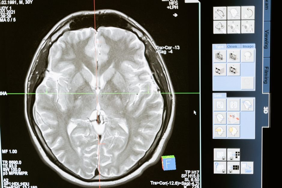
Meanwhile, A breakthrough from a University of Michigan research team, published in Nature Medicine, introduces NeuroVFM. This innovative generalist visual foundation model is specifically designed for neuroimaging, standing apart by learning from an unprecedented volume of real-world clinical data.
The Power of “Health System Learning”
NeuroVFM’s strength lies in its unique training approach, dubbed “health system learning.” Instead of relying on meticulously curated, disease-specific datasets or bottlenecked paired radiology reports, NeuroVFM was trained on an immense dataset:
- 5.24 million clinical MRI and CT volumes.
- Originating from 566,915 studies within the UM-NeuroImages dataset.
- Spanning over two decades of routine patient care at Michigan Medicine.
In practical terms, This method allows the model to learn from the rich, uncurated data generated during normal clinical operations, offering a more robust and generalizable understanding of neuroanatomy.
Unpacking Vol-JEPA: The Self-Supervised Engine
At the heart of NeuroVFM’s training is Vol-JEPA (Volumetric Joint-Embedding Predictive Architecture). This advanced self-supervised, vision-only algorithm is a significant extension of earlier JEPA-style methods (I-JEPA and V-JEPA) adapted for volumetric medical images.
For example, Unlike traditional approaches that reconstruct pixels or require explicit labels, Vol-JEPA predicts representations within a learned latent space. This means:
- No labels are needed.
- No report text is required.
- No voxel decoder is used.
The process involves:
- Tokenization: Each 3D volume is broken down into non-overlapping 4x16x16-voxel patches.
- Context & Target Split: The volume is divided into a small, visible “context” and a larger, masked “target” region.
- Student Encoding: A “student encoder” processes the visible context patches.
- Latent Prediction: A “predictor” then combines these context latents with target position encodings to predict the latents for the masked region.
- Teacher Validation: A “teacher encoder” (an exponential moving average of the student) generates the ground-truth target latents, with gradients stopped to ensure stable learning.
- Loss Minimization: Training minimizes a smooth L1 loss between the predicted and teacher latents, effectively teaching the model to understand shared neuroanatomy by predicting missing parts of the brain.
That said, A key design choice is foreground-focused masking, using precomputed head masks, which encourages the encoder to model relevant anatomical structures rather than background noise.
Impressive Performance Across the Board
NeuroVFM’s performance is compelling. Evaluated across 156 diagnostic tasks (74 MRI, 82 CT), it achieved:
- 92.68 AUROC on CT scans.
- 92.49 AUROC on MRI scans.
Interestingly, Crucially, NeuroVFM surpassed all established baselines on the aggregate endpoint, including models trained on similar data but with different objectives (e.g., report/language supervision or voxel reconstruction). This highlights the superiority of Vol-JEPA’s latent prediction approach.
Furthermore, the model demonstrated remarkable training efficiency, completing a full Vol-JEPA run in fewer than 1,000 GPU hours — over 7 times faster than some 3D baselines and fitting 16 times larger batches at equal memory.
Beyond Diagnosis: Practical Applications
However, NeuroVFM isn’t just for diagnosis; it supports a range of downstream tasks by reusing its frozen visual tokens:
- Report Generation: Paired with Qwen3-14B in a LLaVA-1.5 style (dubbed NeuroVFM-LLaVA), it can generate structured key findings from scans.
- Triage: Findings from NeuroVFM-LLaVA can be fed into a reasoning model to determine acuity levels: unremarkable, routine, or urgent.
- Grounded Predictions: An attention-based MIL pooler maps findings to specific image regions, even without explicit region-level annotations.
- Cross-modal Transfer: A CT-trained probe showed robust performance when applied to MRI, with less than a 5-point AUROC drop.
In a silent, one-week prospective study involving 1,155 cases, NeuroVFM-LLaVA achieved an impressive 92.6% balanced triage accuracy, significantly outperforming GPT-5’s 71.2%. It also proved to be vastly more cost-effective for report inference, being over 24 times cheaper and 23 times less carbon-intensive than GPT-5.
Important Considerations: Strengths and Limitations
Meanwhile, While NeuroVFM represents a significant leap forward, the research team responsibly outlines both its advantages and constraints:
Strengths:
- Learns effectively from uncurated clinical scans without needing explicit report or label supervision.
- Establishes a single, shared latent space across both CT and MRI modalities.
- Demonstrates significantly lower inference cost and carbon footprint for report generation compared to frontier LLMs.
- Maintains consistent performance across different equipment manufacturers, field strengths, and diverse demographic subgroups.
Limitations:
- Its triage sensitivity of 86.5% means that critical findings can still be missed (21 out of 155 in the prospective study). The authors therefore position it as a decision support tool, not an autonomous screening solution.
- The model’s weights are released under a non-commercial license (CC-BY-NC-SA-4.0), and it is not FDA-approved for clinical use.
- Like all AI models, it remains susceptible to dataset, architecture, and objective biases.
- Current results are derived from a single academic health system, suggesting a need for broader validation.
Accessing NeuroVFM
The code for NeuroVFM is available under an MIT license, making it accessible for research and development. While some weights require access approval with an institutional email, the open-source nature facilitates further exploration and integration.
Expert Perspective
A practical read on NeuroVFM starts with neurovfm. That is where the earliest effects are likely to show up if this development keeps building.
What happens next will come down to adoption speed, policy response, and execution quality. That combination could make NeuroVFM a meaningful reference point across jepa.
For decision-makers, the useful lens is not the headline alone but how model changes priorities once organizations have to respond.
Frequently Asked Questions
Why is NeuroVFM important?
Revolutionizing AI in Medical ImagingThe central development is this: Artificial intelligence is rapidly transforming healthcare, yet one significant hurdle remains: access to vast, diverse, and clean clinical data, especially in sensitive areas like neuroimaging.
What impact could NeuroVFM have?
Traditional AI models often struggle because MRI and CT scans contain identifiable patient features, limiting their public availability.
What should readers watch next with NeuroVFM?
This challenge has historically led to underperforming general models in brain-imaging tasks.Meanwhile, A breakthrough from a University of Michigan research team, published in Nature Medicine, introduces NeuroVFM.
How does this relate to neurovfm?
It connects because the article frames neurovfm as one of the clearest areas where the topic may be felt in practice.
Conclusion
Viewed in context, the next round of reactions will matter as much as the initial announcement. In practical terms, NeuroVFM marks a crucial advancement in the application of AI to clinical neuroimaging. By leveraging “health system learning” and the innovative Vol-JEPA architecture, it overcomes significant data challenges, offering robust performance in diagnosis and a suite of practical downstream applications. While it promises to be a powerful decision support tool, its limitations underscore the ongoing need for careful validation and responsible deployment in real-world clinical settings.