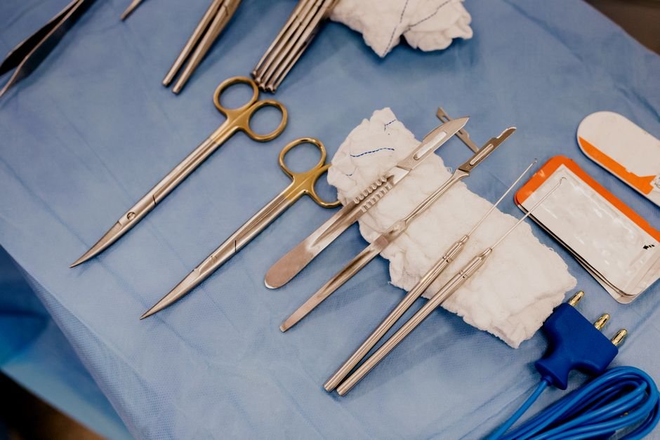
Revolutionizing Surgical Precision with AI
At a glance, Minimally invasive surgeries have transformed modern medicine, offering significant advantages like smaller incisions, reduced pain, and faster recovery times. However, the delicate task of guiding surgical tools through the intricate pathways of the human body, often relying on real-time 2D X-ray images, presents a substantial challenge. This traditional approach demands immense skill and carries inherent risks, as clinicians must mentally translate flat images into a 3D understanding of tool placement.
Table of Contents
- Revolutionizing Surgical Precision with AI
- Introducing xvr: A Patient-Specific AI Solution
- How xvr Works: Tailoring AI to Each Patient
- Transforming Surgical Safety and Accessibility
- The Road Ahead for xvr
- Expert Perspective
- Frequently Asked Questions
- Synthetic X-ray Generation
- Patient-Specific Model Training
- The Foundation Model
- Why is Minimally Invasive Surgery AI important?
- What impact could Minimally Invasive Surgery AI have?
- What should readers watch next with Minimally Invasive Surgery AI?
- How does this relate to patient?
Meanwhile, Existing methods for localizing surgical devices, such as manual alignment of X-rays with preoperative 3D scans like CT or MRI, are often slow and cumbersome. While artificial intelligence has shown promise in streamlining this process, previous AI tools have struggled with robustness across the diverse anatomies of different patients, making them impractical for widespread clinical use.
Introducing xvr: A Patient-Specific AI Solution
A groundbreaking new AI technique, named xvr (X-ray volume registration), developed by scientists and clinicians at MIT and collaborating institutions, is set to overcome these limitations. This innovative system accurately and rapidly matches real-time X-rays captured during surgery with a patient’s unique preoperative 3D medical scan. The impact could be profound: making it significantly easier for clinicians to precisely navigate minimally invasive surgical tools, leading to faster, safer, and more effective procedures.
In practical terms, What sets xvr apart is its remarkable adaptability. Unlike prior AI models that aimed for a one-size-fits-all solution, xvr is designed to tailor itself to each individual patient.
It adapts in approximately five minutes and can then automatically align a patient’s X-rays with their 3D scans in mere seconds, achieving sub-millimeter precision. This level of performance is an order of magnitude better than existing AI methods across a wide spectrum of patients, body parts, and medical procedures.
“A majority of Americans live more than an hour away from a center that can perform noninvasive procedures, like emergency stroke interventions. An hour in stroke time is incredibly substantial. Making these procedures easier by combining 2D and 3D information enables these types of highly specialized life-saving procedures to be more accessible to much broader parts of the population,” says Vivek Gopalakrishnan, lead author of the paper published in Nature and a postdoc at the MIT Computer Science and Artificial Intelligence Laboratory (CSAIL).
How xvr Works: Tailoring AI to Each Patient
For example, The ingenuity of xvr lies in its patient-specific machine learning approach. Instead of attempting to create a universal model, the researchers built a system that deeply specializes for an individual. Here’s a simplified breakdown:
Synthetic X-ray Generation
For a given patient, xvr takes their preoperative 3D scan (MRI or CT) and uses a physics-based simulation to generate thousands of realistic, synthetic X-rays from various angles. This process creates about 1,000 images per second, ensuring the simulated data is highly accurate and free from “hallucinations” often seen in other generative AI.
Patient-Specific Model Training
That said, These simulated data are then used to train an AI model specifically for that patient, enabling it to accurately align their 2D X-rays with their 3D scan in seconds. While training from scratch for each patient would take about 12 hours, making it impractical for emergencies, xvr employs a clever solution.
The Foundation Model
To achieve rapid adaptation, the researchers first pre-trained a more versatile “foundation model.” This model was trained using xvr on whole-body 3D medical scans from over 2,000 diverse patients, generating synthetic X-rays from this vast dataset. This pretrained model can then adapt to a new patient in just about five minutes, delivering the same high accuracy as if it had been trained from scratch.
Interestingly, This innovative two-step process provides the best of both worlds: patient-specific accuracy delivered in a rapid timeframe crucial for emergency medical scenarios.
Transforming Surgical Safety and Accessibility
The implications of xvr are far-reaching. By providing clinicians with a clear, precise 3D understanding of surgical tool placement in real-time, the technique promises to:
- Enhance Safety: Significantly reduce the risk of accidental damage to surrounding tissues.
- Improve Precision: Allow for more accurate navigation, especially in complex procedures like angioplasty or emergency stroke interventions.
- Reduce Training Burden: Make it easier for clinicians to become proficient, potentially shortening the extensive training period currently required to interpret grainy 2D images in a 3D context.
- Increase Accessibility: Broaden access to highly specialized, life-saving procedures by making them less technically challenging and thus more widely available, particularly in areas underserved by specialized medical centers.
The Road Ahead for xvr
However, The research team rigorously tested xvr on the largest available dataset of real 2D/3D registrations, incorporating data from five hospitals covering numerous bones and organ systems in both adult and pediatric patients. Its superior accuracy and robustness, coupled with its speed, confirm its potential for emergency surgeries and even for improving robotic surgery technologies.
Looking to the future, the researchers plan to further optimize xvr for even faster real-time deployment, conduct additional studies to verify its reliability in diverse clinical situations, and extend its capabilities to handle more complex scenarios, such as moving body parts during surgery. Collaborations with surgical robotics companies and clinical groups are already underway to translate this groundbreaking research into practical, life-saving tools.
Expert Perspective
A practical read on Minimally Invasive Surgery AI starts with patient. That is where the earliest effects are likely to show up if this development keeps building.
What happens next will come down to adoption speed, policy response, and execution quality. That combination could make Minimally Invasive Surgery AI a meaningful reference point across procedures.
For decision-makers, the useful lens is not the headline alone but how surgical changes priorities once organizations have to respond.
Frequently Asked Questions
Why is Minimally Invasive Surgery AI important?
Revolutionizing Surgical Precision with AIAt a glance, Minimally invasive surgeries have transformed modern medicine, offering significant advantages like smaller incisions, reduced pain, and faster recovery times.
What impact could Minimally Invasive Surgery AI have?
However, the delicate task of guiding surgical tools through the intricate pathways of the human body, often relying on real-time 2D X-ray images, presents a substantial challenge.
What should readers watch next with Minimally Invasive Surgery AI?
This traditional approach demands immense skill and carries inherent risks, as clinicians must mentally translate flat images into a 3D understanding of tool placement.Meanwhile, Existing methods for localizing surgical devices, such as manual alignment of X-rays with preoperative 3D scans like CT or MRI, are often slow and cumbersome.
How does this relate to patient?
It connects because the article frames patient as one of the clearest areas where the topic may be felt in practice.